
Assess transcranial doppler for cerebral AVMs with MedTech Edge. Review serial measurement limits, probe options and hospital reporting requirements.
Hospital use of transcranial doppler for cerebral AVMs can add selected-vessel blood-flow velocity measurements to a specialist assessment, where clinically appropriate. We supply neurovascular diagnostic equipment through MedTech Edge and help Australian hospital teams evaluate acquisition, monitoring and reporting requirements.
What Can TCD Contribute to an AVM Assessment?
Transcranial Doppler, or TCD, measures blood-flow velocity rather than the complete anatomy of a cerebral arteriovenous malformation. Its findings should be interpreted alongside anatomical imaging and the clinical assessment. A velocity measurement does not by itself establish the AVM’s size, treatment response or future hemorrhage risk.
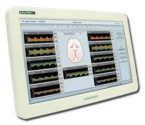
Viasonix’s AVM measurement reference describes velocity and pulsatility patterns that may be assessed using TCD. This manufacturer information explains the application, but it is not a stand-alone clinical protocol or proof that a particular monitoring schedule improves patient outcomes.
Transcranial Doppler for Cerebral AVMs Needs Repeatable Acquisition
For serial measurements, the clinical team should first define the question being investigated and decide whether TCD is appropriate. Comparing examinations requires more than matching a value on two reports. Differences can reflect signal acquisition, physiological conditions or a genuine haemodynamic change.
Clinical teams using transcranial doppler for cerebral AVMs should document:
- Target vessel: Identify the artery, side and acoustic window used for the examination.
- Acquisition settings: Record depth and relevant probe and system settings so later examinations can be compared appropriately.
- Signal quality: Document limitations, uncertain vessel identification and any inadequate acoustic window.
- Patient context: Record relevant physiological and clinical conditions that may affect interpretation.
- Reporting: Retain waveforms and measurements with a consistent description of the examination and its limitations.
Specialist interpretation remains necessary. Repeatable acquisition supports comparison, but it does not make every change clinically significant. Where signal quality is inadequate, the report should state the limitation rather than imply a reassuring result.
Assess Dolphin Equipment Against the Intended Workflow
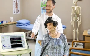
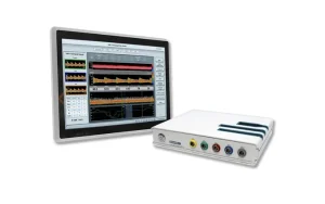
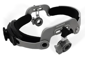
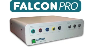
We offer Viasonix Dolphin TCD systems and the Dolphin/XF robotic probe. The robotic option assists vessel searching and monitoring, with a dedicated headset. It should be evaluated for the intended patients, examination duration and staffing model rather than assumed to remove operator oversight.
During evaluation, compare manual and assisted probe positioning, access to saved waveforms and the ease of reviewing prior examinations. Serial assessments across separate appointments and continuous monitoring during one examination are different workflows. State which one your department needs.
Check the proposed system configuration against portability, probe requirements and reporting needs. Dolphin systems offer DICOM and HL7 connectivity features, but hospital integration should be confirmed with your IT team, including interfaces, licensing, patient identifiers and acceptance testing.
Define the Evaluation Before Procurement
Bring clinicians, biomedical engineering and IT into the discussion. Agree the measurement task, operator training requirements, data retention and reporting process before comparing configurations. Ask what sales and after-sales support apply to the proposed equipment and what is included in the quotation.
To evaluate transcranial doppler for cerebral AVMs, request a workflow-based Dolphin demonstration. Include your hospital, clinical evaluation purpose, preferred acquisition method and reporting requirements so we can discuss a configuration for your team to assess.