Pulse Wave Velocity: A New Feature of Viasonix Falcon
04 August 2021
A new feature of pulsed wave velocity (PWV) has been introduced to Viasonix Falcon system. PWV is a simple and non-invasive method to assess arterial stiffness or elasticity. Contraction of the left ventricle generates a pulse wave which is propagated throughout the arterial tree. PWV is obtained by calculation of the pulse wave travel distance between two points in the arterial tree, divided by the pulse wave travel time. Increase of arterial stiffness is believed to be associated with an increase of the propagation speed of the pulse wave in the arteries, i.e., PWV.
During a PWV test, pulse waves at two predefined arterial sites are detected. Most frequently used sites for measurements are the carotid and femoral arteries, brachial and ankle arteries, and the femoral and ankle arteries. The time delay between two arterial sites is automatically determined by a measurement device. The PWV is then calculated based on the distance between two arterial sites where pulse waves are recorded.
The Falcon system has a dedicated PWV protocol which allows users to easily measure faPWV (femoral to ankle PWV) and baPWV (brachial to ankle PWV). The measurement of PWV includes the following steps and takes just a few minutes:
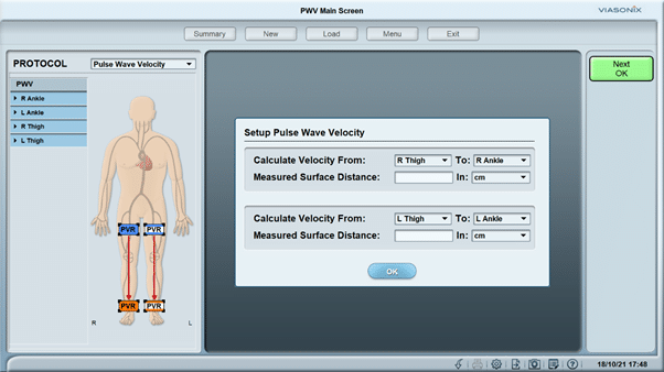
1. Apply blood pressure cuffs to thigh and ankle for faPWV measurement or upper arm and ankle for baPWV measurement and manually enter the distance between the two blood pressure cuffs (Figure 1).
[/vc_column_text][vc_single_image image=”775″ description_text=”Figure 1. Bilateral Setup for Pulse Wave Velocity (PWV).”][vc_column_text]2. Inflate blood pressure cuffs to a venous occlusion pressure (65 mmHg).
3. Pulse Volume Recording (PVR) waveforms at the sites of blood pressure cuffs are displayed on the screen.
4. The Falcon system automatically detects the time delay of the pulse wave between the two blood pressure cuffs.
5. Based on the distance between the two blood pressure cuffs entered by the operator, the Falcon system calculates the PWV automatically (Figure 2).
[/vc_column_text][vc_single_image image=”776″ description_text=”Figure 2. Viasonix Falcon Pro vascular diagnostic system.”][vc_column_text]
The Falcon system allows the PWV measurement to be performed unilaterally or bilaterally. When the PWV test is performed bilaterally, measurements for the right and left sides are carried out simultaneously and does not add any additional time. In this example, faPWV test is performed bilaterally. On the button of the screen, the time delay between the femoral artery and ankle artery (TT), the distance between the femoral artery and ankle artery (Dist.) and faPWV value on each side are displayed (Figure 2).
There have been strong positive correlations reported between cfPWV (carotid to femoral PWV) and baPWV (brachial to ankle PWV), as well as between cfPWV and bfPWV (brachial to femoral PWV). Hence, it is believed that all three parameters of PWV are able to represent central arterial stiffness. Whilst on the other hand, faPWV is considered to represent peripheral arterial stiffness.
In Australia and New Zealand, the Viasonix Falcon system is supplied by MedTech Edge. This state of art vascular diagnostic system is capable of performing a wide range of vascular physiologic tests, including ankle-brachial index (ABI), toe-brachial index (TBI), pulse volume recording (PVR), photoplethysmography (PPG), air plethysmography (APG) and many other tests. To learn more about the Viasonix Falcon system, please contact MedTech Edge in Melbourne.
Optimized by: Netwizard SEO


