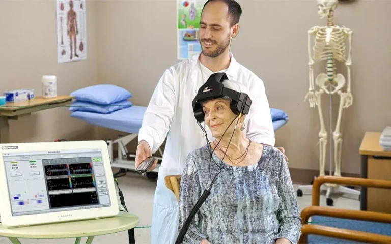
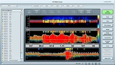
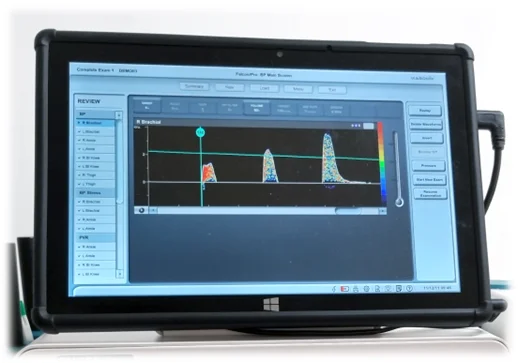
MedTech Edge supports clinicians in Australia with the TCD bubble study. The Dolphin/XF system delivers speed, comfort, and accuracy in PFO screening.
Detecting a patent foramen ovale (PFO) is vital in patients with unexplained stroke, transient ischemic attack (TIA), or other neurological conditions linked to right-to-left shunts. While transesophageal echocardiography (TEE) has long been used for confirmation, it is semi-invasive, time-consuming, and often uncomfortable. The TCD bubble study provides a safe, non-invasive alternative that enables rapid, repeatable screening without the need for sedation or specialised imaging suites.
The TCD bubble study has been performed for decades, but its accuracy and ease of use have often relied on operator skill and the stability of probes. Maintaining consistent signals on both sides of the brain can be challenging, especially during patient manoeuvres such as Valsalva. These limitations have historically reduced efficiency and made reproducibility more difficult.
To overcome these challenges, manufacturers have developed robotic, automated bilateral scanning systems that streamline probe placement and improve signal acquisition. These devices enable clinicians to complete tests more quickly, consistently, and with greater patient comfort. Among them, the Dolphin/XF robotic probe system from MedTech Edge stands out for its speed, ease of use, and targeted design for bubble study procedures.
The Dolphin/XF brings several innovations to the table that make it especially suited to complex functional tests like PFO detection:
• Automated Bilateral Scanning: Robotic probes position and maintain stable contact on both sides of the head, reducing operator dependence and improving reproducibility.
• Rapid Signal Acquisition: Bilateral scanning can be completed in seconds, saving time during contrast injections and minimising patient discomfort.
• Comfort and Stability: The lightweight headset allows patients to be seated, lying down, or even standing, offering flexibility in clinical settings such as ICUs or stroke units.
• Specialty Testing Modes: Dedicated protocols for bubble studies, vasomotor reactivity (VMR), and emboli detection make it easier to run targeted, repeatable assessments.
• Advanced Visualisation: High-resolution depth “heat maps” help clinicians quickly identify optimal signal depths, improving accuracy and interpretation.
Many systems can perform a TCD bubble study, but the Dolphin/XF is recognised as a flagship option for rapid, non-invasive PFO screening. Its robotic bilateral scanning reduces operator dependence and helps deliver consistent, repeatable results. By addressing common challenges such as probe stability and patient cooperation, the Dolphin/XF makes bubble study procedures more reliable and efficient.
The choice of diagnostic equipment depends on factors such as patient volume, workflow, and staff expertise. The Dolphin/XF is designed to meet these needs by combining speed, comfort, and advanced testing protocols in one system. This balance of innovation and practicality allows healthcare providers to strengthen their screening processes and achieve better outcomes for patients.
The TCD bubble study remains an essential tool for early detection of PFO and other right-to-left shunts, providing a fast, patient-friendly alternative to more invasive imaging techniques. With the Dolphin/XF, clinicians can achieve greater efficiency, accuracy, and comfort in their screening process, ultimately supporting better outcomes for patients.
As part of a comprehensive TCD portfolio, the Dolphin/XF reflects how our team at MedTech Edge continues to advance neurological diagnostics — combining innovation with practical solutions for modern healthcare settings.
Contact us at MedTech Edge today to see how we can support your PFO screening and diagnostic programs.
Related Blog Article: Transcranial Doppler (TCD) for Subarachnoid Haemorrhage—Early Vasospasm Alerts for Neurocritical Care

Improve patient outcomes with MedTech Edge in Melbourne through reliable advanced TCD devices for cerebral autoregulation and CO₂ vasomotor reactivity.
Importance of Assessing Cerebral Autoregulation in Critical Care
Impaired cerebral autoregulation places patients with traumatic brain injuries, strokes, or post-surgical complications at greater risk of adverse outcomes. Poor regulation of blood flow can result in either inadequate perfusion or elevated intracranial pressure, both of which may cause additional neurological damage. Early and precise evaluation is essential to prevent secondary injury and improve recovery prospects.
Unstable cerebral perfusion can result in secondary brain injuries that may be preventable with appropriate monitoring. Assessing CO₂ vasomotor reactivity allows healthcare professionals to detect early changes in vascular response, supporting rapid clinical decisions. Such proactive measures significantly enhance patient safety and improve the chances of recovery.
Transcranial Doppler (TCD) is a non-invasive diagnostic tool that measures blood flow velocity within the cerebral arteries. Its real-time feedback enables clinicians to evaluate vascular responses immediately and make informed adjustments to patient care.
TCD remains a preferred method due to its portability and bedside compatibility. Its ability to continuously monitor cerebral circulation provides valuable information without exposing patients to additional risks. Understanding how CO₂ levels influence vessel dilation and constriction is central to the accurate interpretation of the data collected.
Accurate CO₂ vasomotor reactivity testing relies on a structured approach to ensure precise and consistent results. Following these steps helps clinicians optimise cerebral Doppler ultrasound measurements and support informed decision-making in neurocritical care.
• Patient Preparation – Position the patient comfortably and establish baseline readings under stable conditions while ensuring all monitoring systems are accurately calibrated.
• CO₂ Manipulation – Use controlled hyperventilation or administer CO₂-enriched gas to create measurable changes in cerebral blood flow.
• Data Collection – Record mean blood flow velocities with Transcranial Doppler during both baseline and altered CO₂ states for reliable comparisons.
• Interpretation and Assessment – Calculate the vasomotor reactivity index and compare against set thresholds to identify patients at higher risk requiring closer monitoring or intervention.
Continuous monitoring with cerebral Doppler ultrasound provides essential real-time data that guide treatment decisions for critically ill patients. By combining Transcranial Doppler measurements with other modalities, such as intracranial pressure monitoring and multimodal neuromonitoring, clinicians gain a comprehensive understanding of cerebral health.
Featured transcranial Doppler Products from MedTech Edge:
• Dolphin Max – A fully integrated cerebral Doppler ultrasound unit with an internal battery, ideal for mobile use and bedside neurovascular monitoring.
• Dolphin 4D – Offers advanced four-dimensional Doppler imaging for precise vessel visualisation and blood flow assessment.
• Dolphin IQ – A modular TCD component designed to upgrade existing diagnostic setups without needing a full system replacement.
• Dolphin XF – Robot – A robotic bilateral TCD system that automates probe positioning for consistent and continuous dual-hemisphere monitoring.
Combining cerebral autoregulation and CO₂ vasomotor reactivity testing allows clinicians to deliver superior care and reduce risks for vulnerable patients. At MedTech Edge, we provide comprehensive, customisable solutions for advanced neurovascular monitoring. Our range of Transcranial Doppler systems and accessories is designed to meet the diverse needs of hospitals and specialised care units.
Contact our team today to learn how we can support your vascular diagnostic services with innovative and reliable technology.
Related Blog Article: Dolphin TCD Machines for Brain Death Assessment – Available at MedTech Edge for AU & NZ

Support Australian ICU teams with MedTech Edge’s reliable Transcranial Doppler devices for safe and accurate monitoring of traumatic brain injury patients.
Transcranial Doppler for traumatic brain injury provides real-time insights into cerebral blood flow, supporting timely decisions in critical care. This non-invasive tool allows ICU teams to monitor brain function at the bedside without added patient risk. Early detection of blood flow disturbances improves outcomes and guides treatment strategies for complex neurological cases.
Traumatic brain injury disrupts the normal regulation of blood supply to brain tissue, increasing the chance of secondary damage such as ischemia. These disturbances compromise oxygen delivery and worsen neurological outcomes if not addressed promptly. Detecting changes early is vital to reducing the risk of permanent impairment.
ICU teams often struggle to track cerebral perfusion changes accurately using traditional monitoring techniques. Fluctuating intracranial pressures and limited visibility into cerebral hemodynamics make timely intervention difficult. Transcranial Doppler offers a direct, real-time method to assess these changes and guide immediate care.
Transcranial Doppler uses ultrasound technology to measure blood flow velocity within major cerebral arteries. High-frequency sound waves travel through the skull, reflecting off moving red blood cells to generate precise data. This process provides clinicians with continuous feedback on cerebral circulation.
The technique is non-invasive, portable, and safe for repeated use in critical care environments. Clinicians can quickly obtain measurements without moving unstable patients. Parameters such as flow velocity and cerebrovascular resistance offer valuable insight into cerebral function.
At MedTech Edge, we supply advanced TCD devices, including:
• Dolphin Max
• Dolphin 4D
• Dolphin IQ
• Dolphin XF – Robot
Bedside TCD monitoring enhances critical care by providing immediate, accurate data. This supports rapid decision-making and helps prevent secondary brain injuries caused by delayed treatment.
Key benefits include:
• Real-time decision-making: Detect cerebral blood flow changes instantly to guide urgent interventions.
• Non-invasive and repeatable: Monitor patients frequently without introducing additional risks.
• Portability: Perform scans directly at the bedside, reducing the need to move critical patients.
• Guidance for treatment: Guide treatment decisions, medication use, and breathing support.
• Early detection: Identify vasospasm and hypoperfusion before they escalate into severe complications.
Incorporating TCD into ICU protocols improves safety and recovery rates for traumatic brain injury patients by detecting subtle cerebral changes early and preventing secondary complications. This real-time data supports informed clinical decisions and more effective treatments. With proper training and technology adoption, TCD is set to become a standard tool in modern neurocritical care, driving better patient outcomes.
A study in the Asian Journal of Neurosurgery revealed that patients with abnormal TCD readings, such as flow velocities above 120 cm/s or below 35 cm/s, had a 3.87 times higher risk of poor outcomes and a 9.96 times greater risk of mortality, reinforcing the need for precise bedside cerebral flow monitoring in ICU care.
At MedTech Edge, we provide trusted transcranial Doppler solutions to improve traumatic brain injury management. Our bedside monitoring tools help ICU teams act quickly, protect brain function, and enhance recovery. Supported by global partners and proven expertise, we deliver innovative technology that supports exceptional patient outcomes.
Contact us today to explore how our solutions can help transform your critical care practices.
Related Blog Article: Exploring and Comparing the Various Transcranial Doppler Applications in Neurology and Stroke Management

MedTech Edge provides leading peripheral vascular diagnostics in Australia & New Zealand, offering ABI, PVR & Doppler systems for early PAD detection.
Peripheral Artery Disease (PAD) occurs when plaque builds up in the arteries supplying the legs, leading to reduced blood flow and increased cardiovascular risk. This condition is common among older adults but also affects individuals with specific lifestyle and health-related factors. Early warning signs include leg pain during walking, slow-healing wounds on the feet, or numbness and coldness in the lower limbs. Identifying these symptoms early ensures timely referral for further vascular evaluation.
Early diagnosis of PAD depends on reliable vascular testing, which provides clinicians with measurable data beyond physical examination alone. Delayed detection often results in advanced disease requiring surgical intervention, whereas timely screening helps guide conservative treatment strategies. These methods allow practitioners to monitor disease progression and tailor management plans, ultimately reducing the risk of severe complications such as amputation or cardiovascular events.
The value of non-invasive testing in PAD is supported by evidence. The study “Noninvasive physiologic vascular studies: A guide to diagnosing peripheral arterial disease” highlights how physiologic vascular studies such as ABI, Doppler waveforms, and PVR offer reliable insights into arterial function and disease progression (Sibley et al., 2017).
ABI is one of the simplest yet most effective tools in peripheral vascular diagnostics. It compares blood pressure measurements taken at the ankle with those in the arm, providing a reliable indicator of arterial obstruction. Abnormal readings highlight impaired blood flow and support early referral for further imaging.
The main advantages of ABI testing are:
• Non-invasive – Conducted using standard blood pressure cuffs and Doppler probes, with no patient discomfort.
• Quick and efficient – Results are available within minutes, making it suitable for routine screening.
• Reliable baseline tool – Detects early changes in circulation before symptoms escalate.
• Cost-effective – A low-cost method that provides high clinical value in primary care and specialist settings.
PVR testing complements ABI by analysing volume changes in the limb during the cardiac cycle. This approach evaluates the quality of blood flow through waveform interpretation, highlighting arterial narrowing or occlusion. When combined with ABI, PVR enhances diagnostic accuracy, offering clinicians a broader understanding of circulatory health in the lower extremities.
Doppler ultrasound adds a real-time imaging dimension to peripheral vascular diagnostics. Using sound waves to map arterial flow enables clinicians to detect narrowing or blockages with precision. Unlike pressure-based tests, Doppler provides direct visual evidence of the severity and location of disease.
The primary benefits include:
• Real-time imaging – Offers immediate insights into arterial conditions.
• Detailed localisation – Pinpoints the exact site of narrowing or blockage.
• Non-invasive and safe – No radiation or invasive procedure required.
• Supports treatment planning – Informs whether surgical or non-surgical intervention is required.
• Effective for follow-up – Tracks changes in arterial health after treatment or lifestyle modification.
Accurate PAD screening through ABI, PVR, and Doppler ultrasound enables earlier intervention, reducing complications and improving patient outcomes. At MedTech Edge, we provide advanced peripheral vascular diagnostics with Falcon systems that are comprehensive, adaptable, and designed for diverse clinical needs. Our ultramodern non-invasive technologies make vascular screening precise, scalable, and clinically reliable.
Contact us today to enhance your diagnostic service.
Related Blog Article: Pulse Volume Recording (PVR): Improve Non-Invasive PAD Diagnosis with the Falcon

Enhance neurocritical care with MedTech Edge in Australia, offering advanced Transcranial Doppler platforms for timely vasospasm detection in SAH.
Subarachnoid haemorrhage (SAH) represents one of the most severe neurological emergencies, often caused by a ruptured cerebral aneurysm. Among its complications, delayed cerebral ischaemia from vasospasm poses the greatest threat to survival and long-term recovery. Transcranial Doppler has emerged as a reliable, non-invasive tool that allows real-time monitoring of cerebral circulation, enabling early detection of flow changes and timely intervention to prevent irreversible damage.
TCD is an ultrasound-based technique that measures blood flow velocity within the cerebral arteries. Unlike static imaging modalities, it allows dynamic assessment and can be performed repeatedly at the bedside, making it invaluable in intensive care environments.
At MedTech Edge, we offer tailored TCD solutions designed to provide clinicians with clarity, speed, and diagnostic confidence:
• Dolphin Max: Provides high-resolution imaging for detailed analysis of cerebral haemodynamics, supporting precise interpretation of subtle flow changes.
• Dolphin 4D: Delivers four-dimensional imaging for a comprehensive evaluation of intracranial blood flow, offering deeper insights into vascular dynamics.
• Dolphin IQ: Integrates intelligent algorithms that enhance diagnostic accuracy through automated data interpretation, streamlining clinician decision-making.
• Dolphin XF-Robot: Utilises robotic precision to standardise examinations, minimising operator variability and ensuring consistent, reproducible results that improve patient outcomes.
These advanced platforms empower clinicians with tools to detect, monitor, and respond rapidly to vasospasm risk in SAH patients.
While immediate mortality from SAH is a primary concern, survivors face the ongoing risk of cerebral vasospasm. This delayed narrowing of arteries reduces cerebral blood flow and can lead to secondary ischaemic injury. Detecting vasospasm before infarction occurs is essential to improving survival rates and reducing disability.
Transcranial Doppler provides clinicians with an immediate, bedside tool to identify vasospasm before clinical deterioration occurs. This early detection ensures that treatment can begin at the most critical window, reducing the chance of permanent neurological damage. By delivering continuous, real-time data, TCD allows healthcare teams to make evidence-based decisions with greater confidence.
In the review “Transcranial Doppler Monitoring in Subarachnoid Hemorrhage” (J Neurosonol Neuroimag, 2022; 14[1]: 1–9), Park et al. emphasize that TCD enables real-time, bedside detection of cerebral vasospasm and serial monitoring of treatment efficacy in SAH patients. This evidence underscores TCD’s value as a non-invasive, dynamic monitoring tool that enhances timely intervention in neurocritical care settings following subarachnoid haemorrhage.
The integration of TCD into routine neurocritical workflows has transformed SAH monitoring. Its role goes beyond vasospasm detection and contributes to comprehensive patient care:
• Continuous, real-time cerebral flow assessment
• Non-invasive, bedside application
• Guidance for therapy adjustments
• Objective data to support decision-making
• Improved survival and recovery rates
At MedTech Edge, we are committed to delivering innovative Transcranial Doppler solutions that exceed industry standards and integrate seamlessly into routine clinical practice. By enabling early vasospasm detection in subarachnoid haemorrhage, our products empower neurocritical care teams and reflect our vision to provide clinicians with technologies that genuinely improve patient outcomes.
Take the next step in advancing patient care. Contact us at MedTech Edge today to learn how our TCD solutions can support your neurocritical care team.
Related Blog Article: Transcranial Doppler Machines for Sickle Cell Disease – Early Stroke Detection in Children


